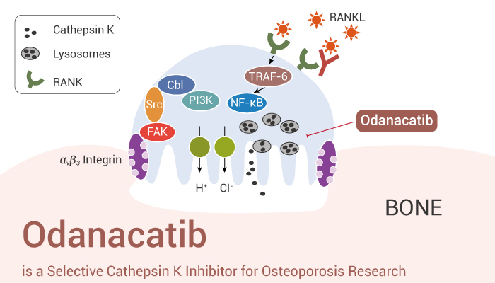
Cathepsin K is a cysteine protease member of the lysosomal protease family of cathepsin. Besides, cathepsin K is the only tissue protease that is highly expressed in osteoclasts. Cathepsin K is a cysteine protease similar to papain, with high matrix degradation activity. Moreover, cathepsin K is the most effective mammalian collagenase. Furthermore, cathepsin K secretes from osteoclasts to the sealed osteoclast bone cell interface, leading to effective degradation of type I collagen. Research has shown that its collagen solubilizing effect requires the binding of chondroitin or sulfated keratin with proteases. Meanwhile, cathepsin K is a recognized component in osteoclasts and plays an important role in the function of osteoclasts and the degradation of protein components in cells. Nonetheless, the lack of tissue protease K activity in the human body can lead to dense bone hyperplasia, characterized by increased bone mineral density and fractures. Here, we will introduce a selective inhibitor of cathepsin K, Odanacatib.
Odanacatib is a Selective Cathepsin K Inhibitor for Osteoporosis Research.
First of all, Odanacatib (MK-0822) is a selective inhibitor of cathepsin K, with an IC50 of 0.2 nM for human cathepsin K. Importantly, Odanacatib is a weak inhibitor of antigen presentation, measured in a mouse B cell line (IC50=1.5±0.4 μM). Particularly, Odanacatib also shows weak inhibition of the processing of the MHC II invariant chain protein Iip10 in mouse splenocytes.
In the second place, Odanacatib dose-dependently reduces CTx release with an IC50=9.4±1.0 nM. Obviously, Odanacatib treated OC accumulates labeled degraded bone matrix proteins in CatK containing vesicles.
Last but not the least, Odanacatib with 30 mg/kg by orally once daily persistently suppresses bone resorption markers and serum bone formation markers. Interestingly, Odanacatib displays compartment-specific effects on trabecular versus cortical bone formation.
All in all, Odanacatib (MK-0822) is a potent and selective inhibitor of cathepsin K and has the potential for osteoporosis research.
References:
[1] Jacques Yves Gauthier, et al. Bioorg Med Chem Lett. 2008 Feb 1;18(3):923-8.
[2] Leung P, et al. Bone. 2011 Oct;49(4):623-635.